The Brain’s “Bravery Cells” Encourage Risky Behavior
Cells in the hippocampus help determine whether to be apprehensive in stressful situations, and they could be stimulated to treat anxiety
:focal(1192x885:1193x886)/https://tf-cmsv2-smithsonianmag-media.s3.amazonaws.com/filer/3a/df/3adf9c08-3c60-4dc4-af03-c1df144704b8/istock-477586286.jpg)
You’re walking alone in the woods when, suddenly, you spy a wolf. You scramble behind a tree and peer out from behind the trunk. Even from a distance you swear you can see the animal’s incisors glint, but it’s far enough away that it doesn’t seem to notice you. Do you ignore the wolf and continue on your way, or stay put?
According to new research, your reaction may have less to do with logically analyzing the situation and more to do with how so-called “bravery cells” in your brain light up in response to the threat. Our brains have been primed from the early stages of evolution to respond to risk in order to keep us safe, but not all risky scenarios are as severe as a hungry wolf in the woods—and sometimes our minds flood us with apprehension when there is no risk at all.
Scientists from Uppsala University in Sweden and Federal University of Rio Grande do Norte in Brazil have pinpointed one thread of the cognitive web that controls anxiety: oriens lacunosum-moleculare interneurons, or OLM cells. These brain cells activate to let us know we are safe in risky situations, and they might provide a new method to counter the debilitating effects of anxiety disorders.
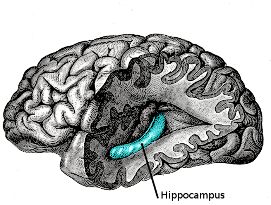
OLM cells are found in the hippocampus, a tiny chunk of tissue smack dab in the middle of the brain that is best known for its role committing short-term to long-term memory. More specifically, OLM cells live in the ventral hippocampus, which crests along the inside of the seahorse-shaped cerebral section. While the opposing dorsal hippocampus lights up during spatial cognitive functions, the ventral hippocampus has been linked to emotions including anxiety.
“Just in the last ten years, scientists have started appreciating the difference between ventral and dorsal hippocampus,” says Sanja Mikulovic, primary author of the new study in Nature Communications and a postdoctoral researcher at Uppsala University. “When we started investigating, we saw different activity that is related to emotional information processing appeared in ventral hippocampus.”
The key to separating these two regions and their functions is measuring their vibrations. Our brains produce waves with a range of frequencies that dictate our thoughts and actions. (Neuroscientists know a lot about how these vibrations happen, their structure and chemistry, but significantly less about why.) Type 1 theta waves, which are higher frequency, have been shown to ripple through the dorsal hippocampus when an animal is moving and exploring. In contrast, lower-frequency type 2 theta waves appear in the ventral hippocampus during stressful situations, such as encountering a predator.
Though both types of theta waves are prevalent in the hippocampus, they occupy unique circuits in the dorsal and ventral sections. Imagine you’re trying to find your way home from work. In that moment, Mikulovic says, Type 1 theta waves surge through the dorsal hippocampus to pull up a spatial map of your route home. But if you see a strange and threatening animal crossing the road, type 2 theta waves would simultaneously appear in the ventral hippocampus. In order to decide whether to continue or turn back, the two types of theta activity interact with one another and influence your decision.
The generation of two different theta waves was once thought to be caused by a certain neurotransmitter, acetylcholine, as well as that molecule's sensitivity to anesthetics. When research debunked this theory, Mikulovic and her colleagues began wondering if the different vibrations came from the cells that produced the waves. The researchers decided to target OLM cells, which had previously been linked to anxiety responses.
The team used a technique called optogenetic activation, which triggers light-sensitive neurons using different colored fiber optics inserted into the brains of mice. Mikulovic and her team found that activating OLM cells increased type 2 theta wave generation in the hippocampus, and inhibiting the cells decreased such activity. The OLM cells, it appeared, were making waves in the brain.
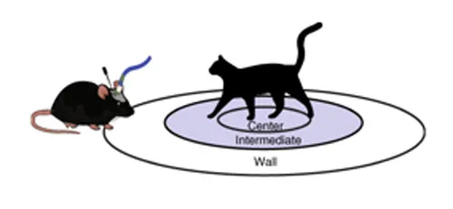
The researchers were also able to link type 2 theta generation to increased risk-taking behavior in response to anxiety-inducing situations. The researchers put the mice in a circular arena with a smelly clump of cat hair at the center. Mice that had their OLM cells stimulated were more likely to explore closer to the center, while mice that had their OLM cells inhibited stayed fearfully at the peripheries.
The results are promising, but as with everything in the brain, there are more nuances to explore. In other studies, type 2 theta waves have been shown to appear in male animals in the presence of female subjects, indicating that theta 2 waves might not be unique to anxiety.
“Is [the mouse] anxious or attracted?” Mikulovic wonders. “We don’t exclude the possibility that there are more subtypes of theta 2 themselves. We want to understand how theta 2 relates to different behaviors.”
Like emotions themselves, the brain is complex and largely ineffable. A single moment causes many different parts of the brain, each with their own function, to activate and interact. Understanding what each part contributes can help us understand how we perceive the world and more effectively control our reactions to those perceptions.
Connecting the OLM cells to theta 2 waves helps elucidate how the hippocampus interacts with other parts of the brain to generate a response to anxiety. The ventral hippocampus has been shown to interact frequently with the prefrontal cortex and the amygdala, which play important roles in decision making. The simple amygdala (metaphorically called the “lizard brain”) generates autonomic fear responses, while the higher cognitively-functioning prefrontal cortex helps make decisions in the face of fearful stimuli, inhibiting the amygdala when need be. Type 2 theta waves likely help synchronize the ventral hippocampus with these regions by literally getting them on the same wavelength.
“The hippocampus communicates with both of those and then sends certain kinds information to help make the decision about whether to be afraid or not,” says Joshua Gordon, director of the National Institute of Mental Health. “We have already discovered that when there is an anxiety provoking stimulus in the world, we tend to see increases in the ability of [theta 2] in the hippocampus to synchronize with [theta 2] in the other structures.”
Anxiety disorders have been linked to a disrupted connection between the prefrontal cortex and the amygdala, and now that the researchers know that OLM cells produce type 2 theta waves, they could carve new pathways for treating anxiety. Like all cells, OLM cells have their own unique sets of receptors and sensitivities that could be manipulated in order to increase theta 2 waves and quell inhibitive or inappropriate anxiety reactions. According to Gordon, there are currently two primary ways to treat anxiety: drugs that bind receptors all over the brain and psychotherapy to teach the prefrontal cortex how to restrain the amygdala. A potential third avenue could be a drug designed to target receptors in the OLM cells to activate type 2 theta waves when anxiety feels unmanageable.
But Gordon warns against sloppy solutions. As of yet, the studies have only been conducted on mice, so there is no definitive proof that the findings are directly applicable to humans. He also points out that research shows that OLM cells are sensitive to nicotine (which is especially enlightening to those of us who chain smoke to cope with anxiety), but smoking should not be considered a long-term solution for treating anxiety because of the addictive properties and other nasty side effects.
“Developing a better nicotine for anxiety isn’t going to lead us down new pathways,” Gordon laughs. “But it does begin to say how we might treat OLM cells.”
In the ever-chugging cognitive engine of the brain, well-oiled OLM cells are able to determine when it’s safe to trudge through the dangerous and the unfamiliar. But even if our brains follow the same basic blueprints, every brain behaves a bit differently. When OLM cells misfire, our brains can panic, even when the perceived threat is completely surmountable. By identifying the role of each cellular cog in the machine, scientists might be able to address these glitches and help our brains run a little smoother.