An Artificial Ear Built By a 3D Printer and Living Cartilage Cells
Cornell scientists used computerized scanning, 3D printers and cartilage from cows to create living prosthetic ears
/https://tf-cmsv2-smithsonianmag-media.s3.amazonaws.com/filer/20130221091140ear-sample-small.jpg)
3D printing is big news: During his State of the Union speech, President Obama called for the launch of manufacturing hubs centered around 3D printing, while earlier this week, we saw the birth of one of the most playful applications of the technology yet, the 3D Doodler, which lets you draw solid plastic objects in 3 dimensions.
Yesterday, Cornell doctors and engineers presented a rather different use of the technology: a lifelike artificial ear made of living cells, built using 3D printing technology. Their product, described in a paper published in PLOS ONE, is designed to help children born with congenital defects that leave them with underdeveloped outer ears, such as microtia.
The prosthesis—which could replace previously used artificial materials with styrofoam-like textures, or the use of cartilage tissue harvested from a patient’s ribcage—is the result of a multistep process.
First, the researchers make a digital 3D representation of a patient’s ear. For their prototype, they scanned healthy pediatric ears, but theoretically, they might someday be able to scan an intact ear on the other side of a patient’s head—if their microtia has only affected one of their ears—and reverse the digital image, enabling them to create an exact replica of the healthy ear.
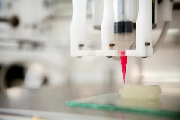
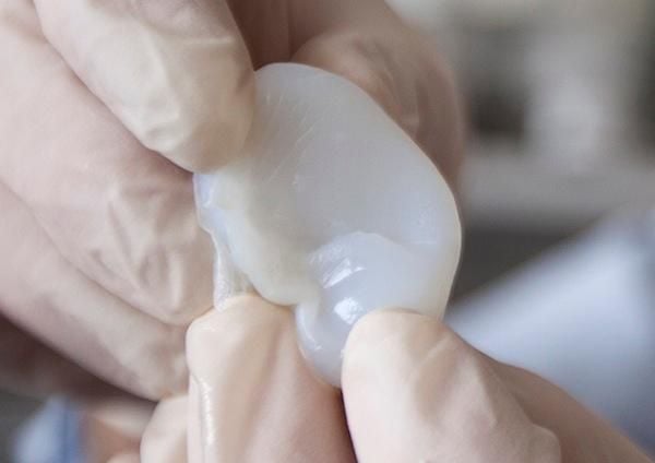
Next, they use a 3D printer to produce a solid plastic mold the exact shape of the ear and fill it with a high-density collagen gel, which they describe as having a consistency similar to Jell-O.


After printing, the researchers introduce cartilage cells into the collagen matrix. For the prototype, they used cartilage samples harvested from cows, but they could presumably use cells from cartilage elsewhere on the patient’s own body in practice.
Over the course of a few days in a petri dish filled with nutrients, the cartilage cells reproduce and begin to replace the collagen. Afterward, the ear can be surgically attached to a human and covered with skin, where the the cartilage cells continue to replace the collagen.
So far, the team has only implanted the artificial ears underneath the skin on the backs of lab rats. After 3 months attached to the rats, the cartilage cells had replaced all the collagen and filled in the entire ear, and the prosthetic retained its original shape and size.
In a press statement, co-author Jason Spector said that using a patient’s own cells would greatly reduce the chance of the body rejecting the implant after surgery. Lawrence Bonassar, another co-author, noted that in addition to congenital defects, the prosthesis could also be valuable for those who lose their outer ear as a result of cancer or an accident. If used for a child with microtia, the ear won’t grow along with the head over time, so the researchers recommend waiting to implant one of their prostheses until the patient is 5 or 6 years old, when ears have normally grown to more than 80 percent of their adult size.
The biggest advantage of the new technology over existing methods is the fact that the production process is customizable, so it could someday produce remarkably realistic-looking ears for each patient on a rapid timescale. The researchers have actually sped up the process since conducting the experiments included in the study, developing the ability to directly print the ear using the collagen as an “ink” and skip making the mold.
There are still a few problems to tackle, though. Right now, they don’t have the means to harvest and cultivate enough of a pediatric patient’s own cartilage to build an ear, which is why they used samples from cows. Additionally, future tests are needed to prove that surgical implantation is safe for humans. The team says they plan to address these issues and could be working on the first implant of such an ear in a human as soon as 2016.
/https://tf-cmsv2-smithsonianmag-media.s3.amazonaws.com/accounts/headshot/joseph-stromberg-240.jpg)
/https://tf-cmsv2-smithsonianmag-media.s3.amazonaws.com/accounts/headshot/joseph-stromberg-240.jpg)