This Year’s Best Photographs Taken Through the Lens of a Microscope

Who knew a turtle’s retina could be so beautiful?
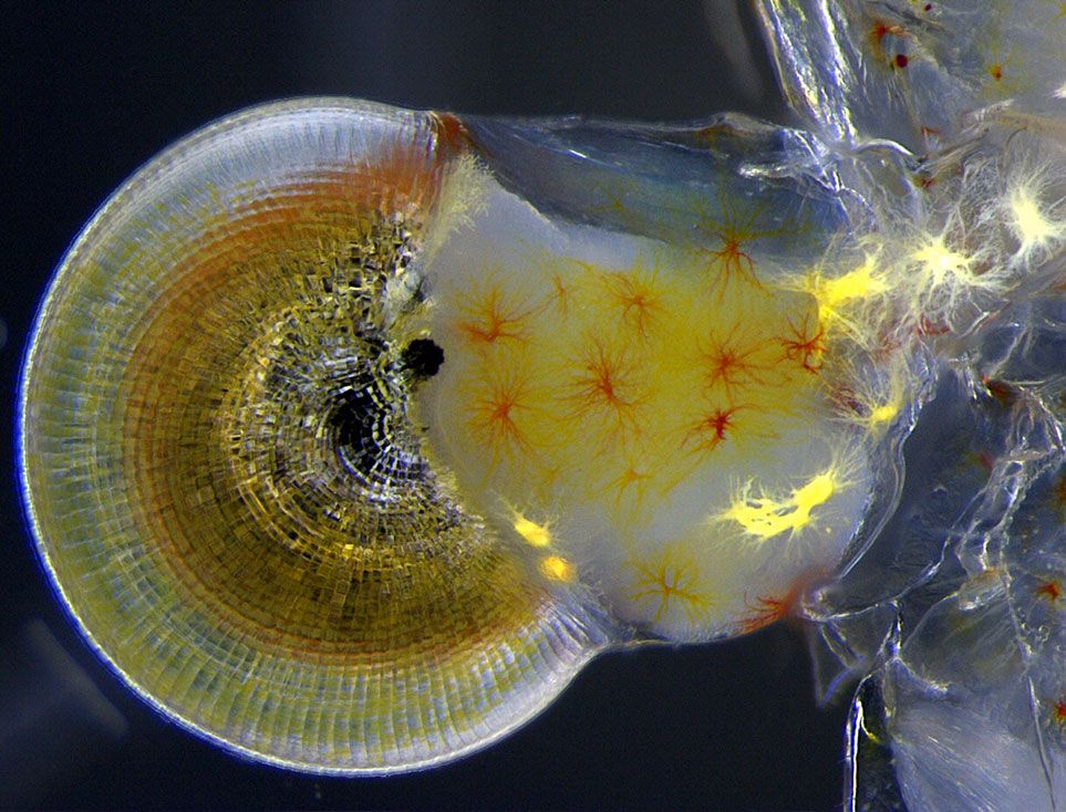
11th Place: Macrobrachium shrimp (ghost shrimp) eye. Vitoria Tobias Santos, Universidade Federal do Rio de Janeiro, Rodrigo Evo Devo Group, Macaé, Rio de Janeiro, Brazil Nikon Small World
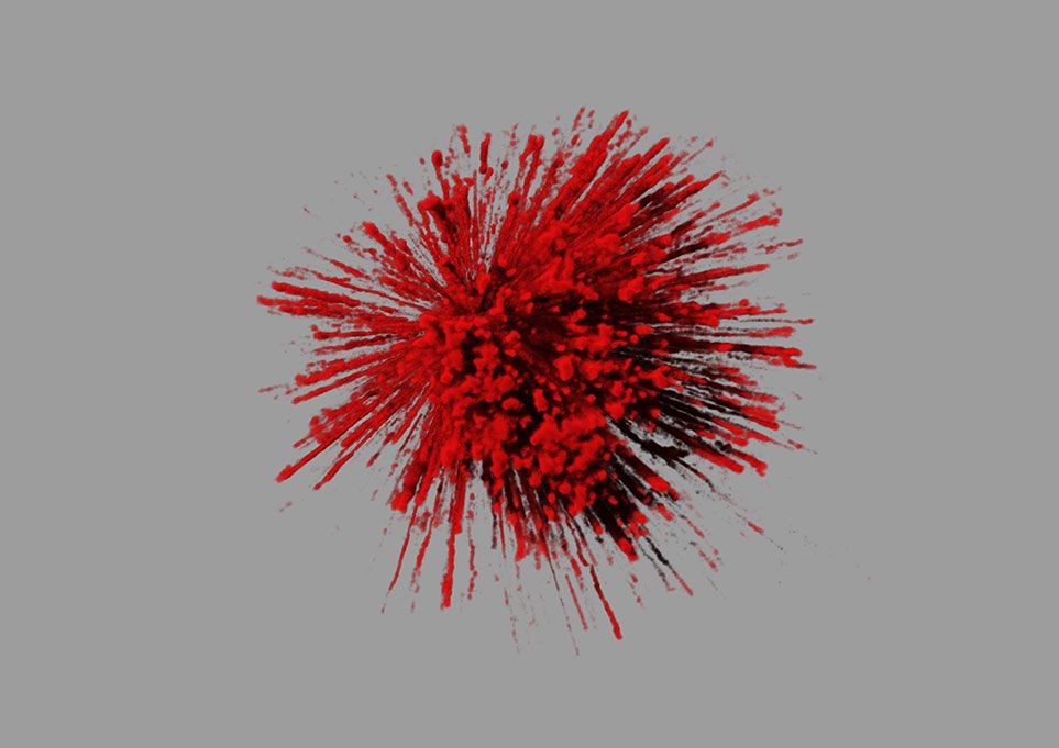
20th Place: The explosive dynamics of sugar transport in fat cells. James Burchfield, The Garvan Institute, Sydney, New South Wales, Australia Nikon Small World
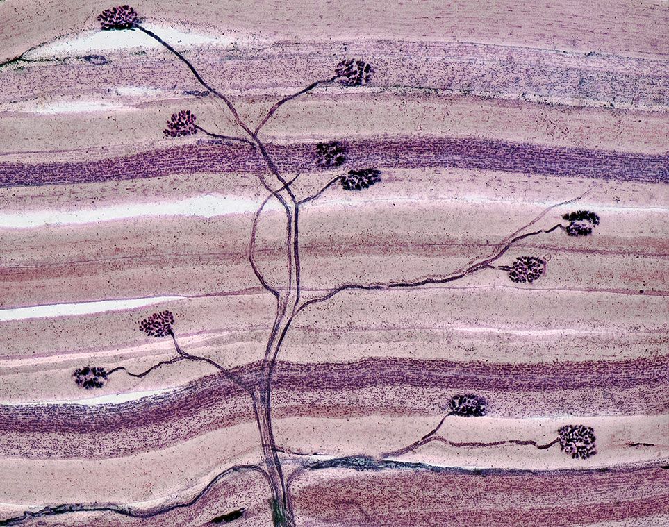
19th Place: Nerve and muscle thin section. David Ward, www.dgward.com, Oakdale, California, USA Nikon Small World
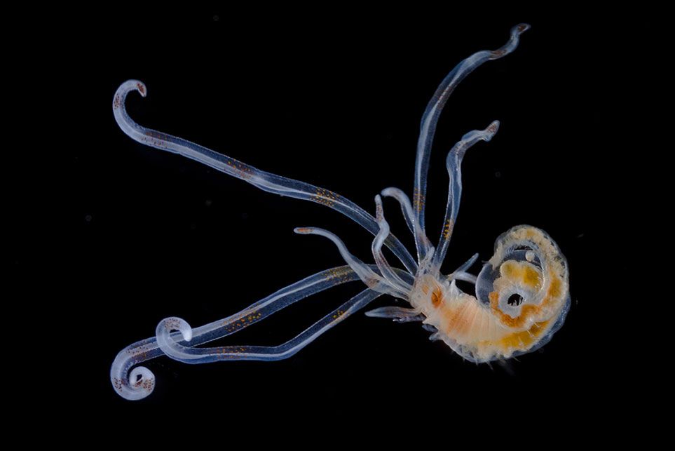
18th Place: Annelid larva. Christian Sardet, Centre National de la Recherche Scientifique, Department of Life Sciences, Strasbourg, France Nikon Small World
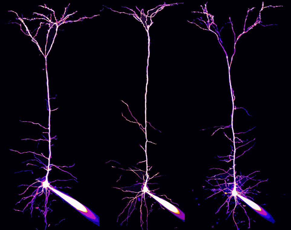
17th Place: Pyramidal neurons and their dendrites visualized in the visual cortex of a mouse brain. Alexandre William Moreau, University College London, Institute of Neurology, London, UK Nikon Small World
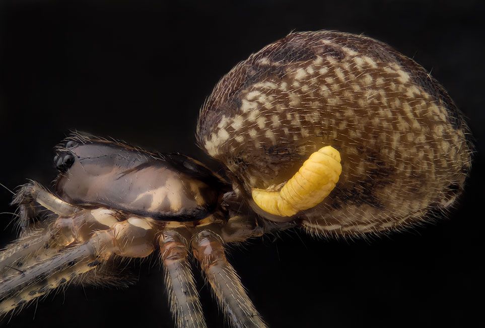
16th Place: Pityohyphantes phrygianus (sheet weaver spider) with a parasitic wasp larva on the abdomen. Geir Drange, Asker, Norway Nikon Small World
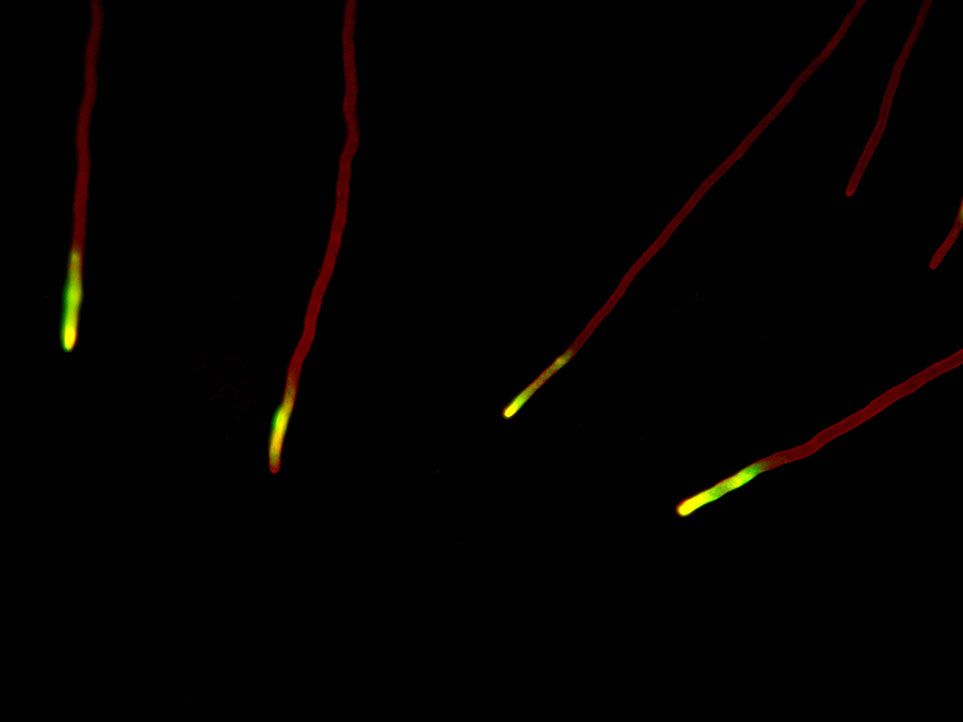
15th Place: Podospora anserina (fungus) filamentous tip cells. Christian Q. Scheckhuber, Goethe University, Frankfurt, Germany Nikon Small World
14th Place: Peripheral nerves in E11.5 mouse embryo. Zhong Hua, Johns Hopkins School of Medicine, Department of Molecular Biology & Genetics, Baltimore, Maryland, USA Nikon Small World
13th Place: Mouse vertebra section. Michael Paul Nelson and Samantha Smith, University of Alabama at Birmingham, Department of Pathology/Neuropathology, Birmingham, Alabama, USA Nikon Small World
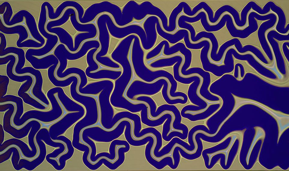
12th Place: Silicon dioxide on polydimethylglutarimide-based resist. Pedro Barrios-Perez, National Research Council of Canada, Information and Communication Technologies, CPFC (Nanofabrication), Ottawa, Ontario, Canada Nikon Small World
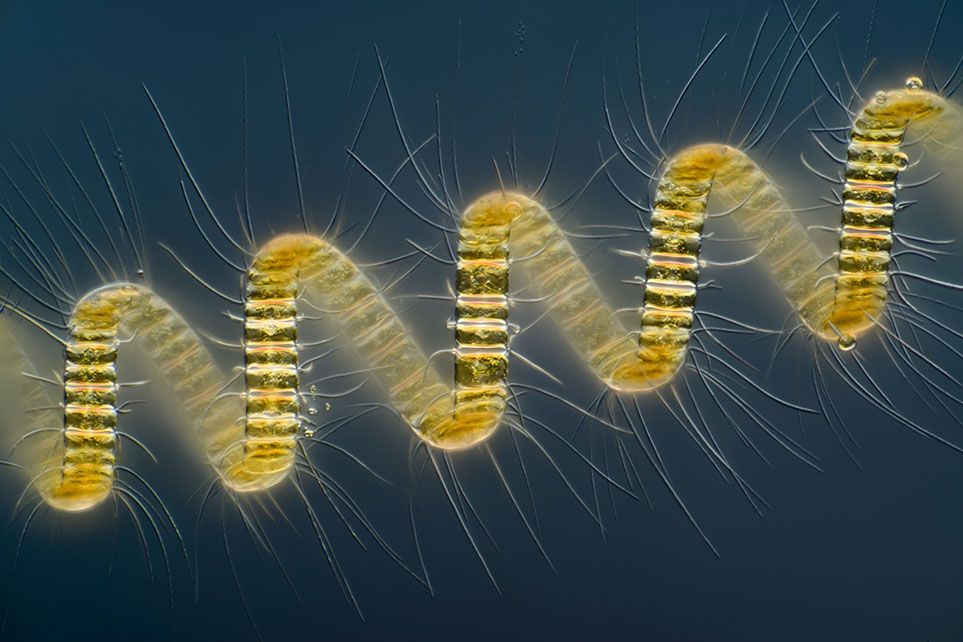
Looking through a microscope can be an awe-inspiring experience. So, why can't photographs of magnified paramecia, neurons and chameleon embryos be celebrated as art? Above: 1st Place Chaetoceros debilis (marine diatom), a colonial plankton organism. Wim van Egmond, Micropolitan Museum, Rotterdam, The Netherlands Nikon Small World
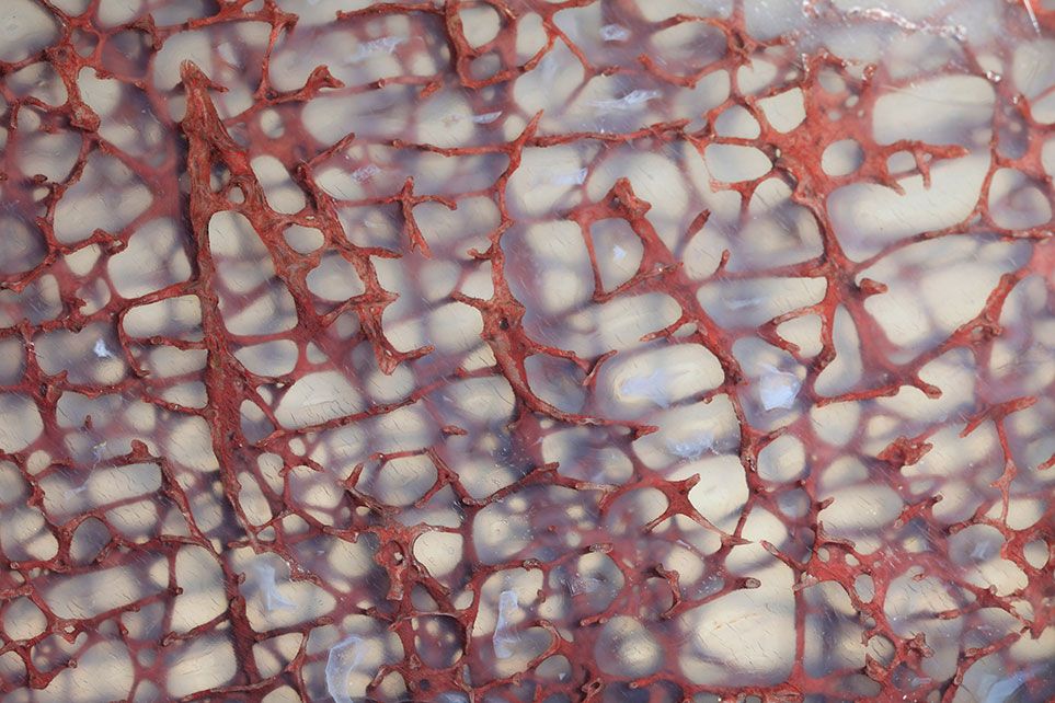
10th Place: Thin section of a dinosaur bone preserved in clear agate. Ted Kinsman, Rochester Institute of Technology, Department of Imaging and Photo Technology, Rochester, New York, USA Nikon Small World
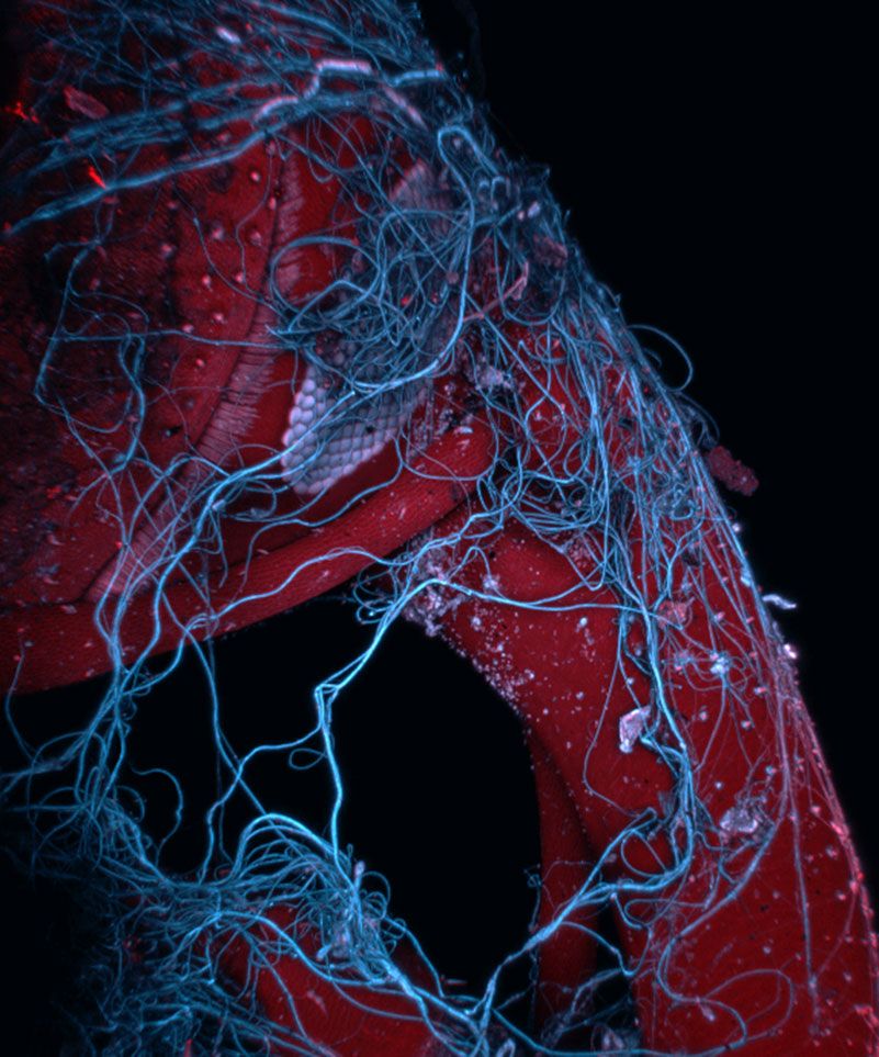
9th Place: Insect wrapped in spider web. Mark A. Sanders, University of Minnesota, University Imaging Centers, Minneapolis, Minnesota, USA Nikon Small World
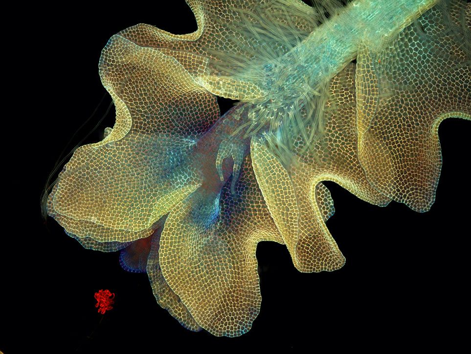
8th Place: Barbilophozia sp. (a leafy liverwort, bryophyte plant) and cyanobacteria. Magdalena Turzanska, University of Wrocław, Institute of Experimental Biology, Department of Plant Developmental Biology, Wrocław, Poland Nikon Small World
7th Place: Adhesive pad on a foreleg of Coccinella septempunctata (ladybird beetle). Jan Michels, Christian-Albrechts-Universität zu Kiel, Institute of Zoology, Functional Morphology and Biomechanics, Kiel, Germany Nikon Small World
6th Place: Chamaeleo calyptratus (veiled chameleon), embryo showing cartilage (blue) and bone (red). Dorit Hockman, University of Cambridge, Cambridge, UK Nikon Small World
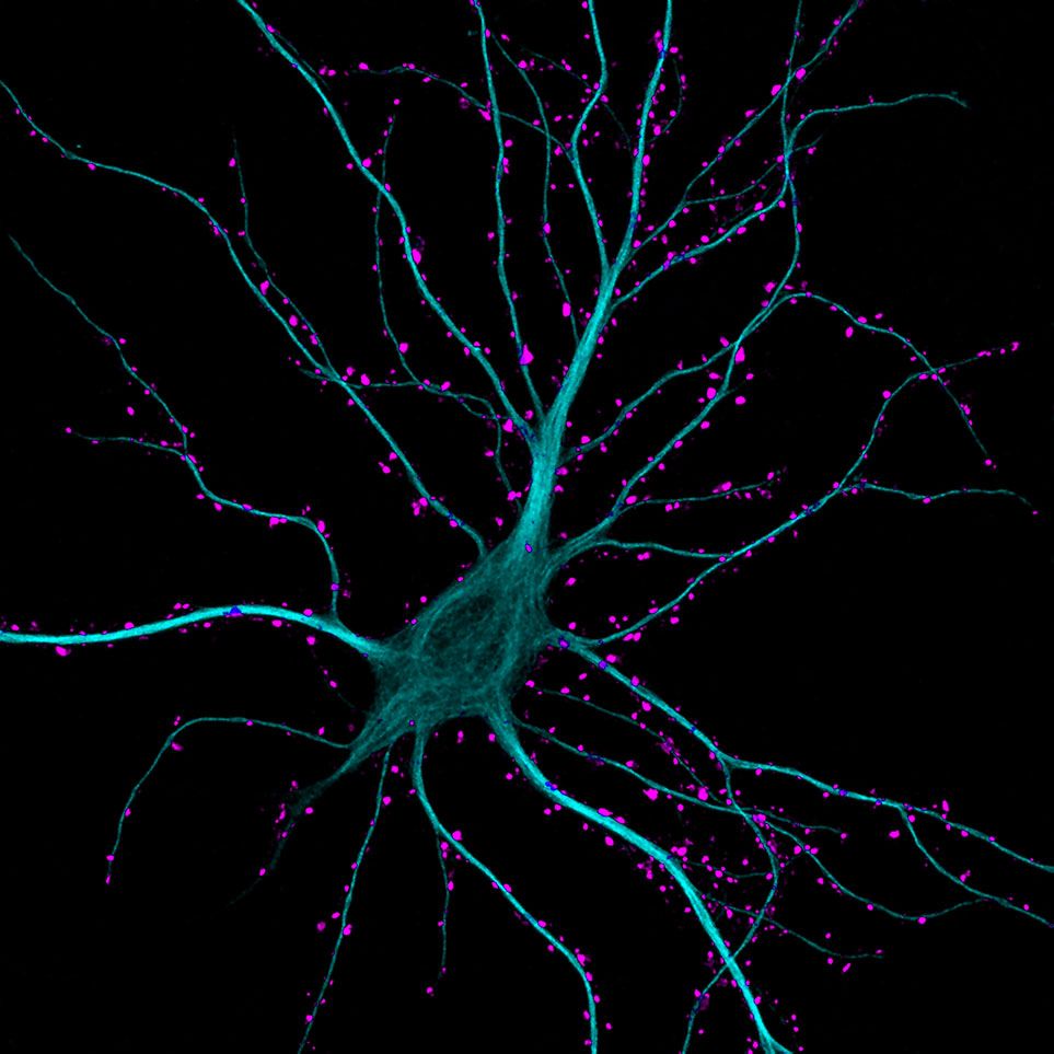
5th Place: Hippocampal neuron receiving excitatory contacts. Kieran Boyle, University of Glasgow, Institute of Neuroscience and Psychology, Glasgow, Scotland, UK Nikon Small World
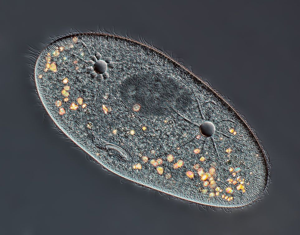
4th Place: Paramecium sp. showing the nucleus, mouth and water expulsion vacuoles. Rogelio Moreno Gill, Panama City, Panama Nikon Small World
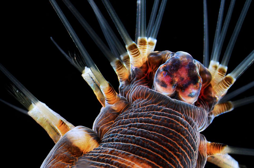
3rd Place: Marine worm. Alvaro Esteves Migotto, Universidade de São Paulo, Centro de Biologia Marinha, São Paulo, Brazil Nikon Small World
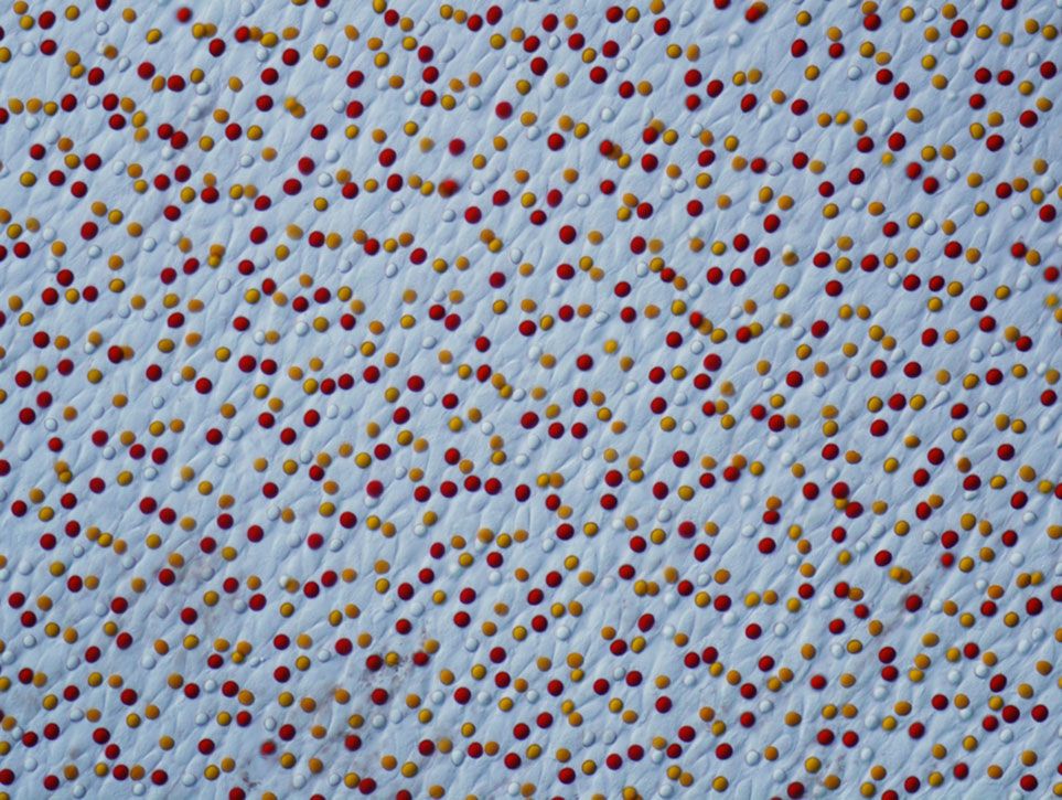
2nd Place: Chrysemys picta (painted turtle) retina. Joseph Corbo, Washington University School of Medicine, St. Louis, Missouri, USA Nikon Small World

Since 1974, Nikon has played host to an annual Small World Competition, aiming to highlight the year's best in photomicrography. In 2013, scientists and professional photographers from more than 80 countries submitted some 2,000 images to the contest. A panel of judges selected the top 20 finishers based on established criteria. According to the competition's website, each photomicrograph had to not only be "a technical document that can be of great significance to science or industry" but also "an image whose structure, color, composition and content is an object of beauty, open to several levels of comprehension and appreciation."

A veteran photomicrographer, Wim van Egmond has entered numerous images for consideration in the last decade, nabbing a total of 20 finalists. For his first prize winner this year, he captured a coil-shaped marine plankton, collected from the North Sea, by stacking more than 90 separate images.

"I approach micrographs as if they are portraits. The same way you look at a person and try to capture their personality, I observe an organism and try to capture it as honestly and realistically as possible," says Egmond in a press release. "At the same time, this image is about form, rhythm and composition. The positioning of the helix, the directions of the bristles, the subdued colors and contrast all bring together a balance that is both dynamic and tranquil."

Take a gander at the rest of the 2013 finalists.

Get the latest Science stories in your inbox.