What Does A Bee Look Like When It’s Magnified 3000 Times?
Photographer Rose-Lynn Fisher uses a powerful microscope to capture all of a bee’s microscopic structures and textures in stunning detail
/https://tf-cmsv2-smithsonianmag-media.s3.amazonaws.com/filer/Collage-Bee-closeup.jpg)
You’ve probably seen a bee fly by hundreds of times in your life, if not thousands. When it arrived, maybe attracted by something you were eating or drinking, you likely shooed it away, or perhaps remained entirely still to avoid provoking a sting.
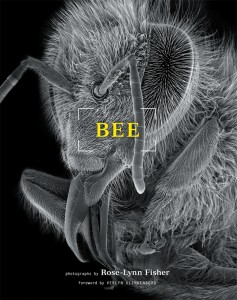
Cover of Bee, a collection of photographs by Rose-Lynn Fisher. Image courtesy Princeton Architectural Press
One thing you probably didn’t do was consider how the bee would look under intense magnification, blown up to 30, 300 or even 3,000 times its original size. But—as photographer Rose-Lynn Fisher has discovered over the past two decades working with powerful scanning electron microscopes (SEMs) to capture images of the insects in remarkable detail—everyday bees feature incredible microscopic structures.
“Once you scratch the surface, you see there’s a whole world down there,” says Fisher, who published her photos in the 2010 book Bee and is having them featured in the new exhibition Beyond Earth Art at Cornell University in January. “Once I started, it became a geographical expedition into the little body of the bee, with higher and higher magnifications that took me deeper and deeper.”
Fisher began creating the images back in 1992. “I was curious to see what something looked like under a scanning electron microscope, and a good friend of mine was a microscopist, and he invited me to bring something to look at,” she says. “I’ve always loved bees, and I had one that I found, so I brought it in to his lab.”
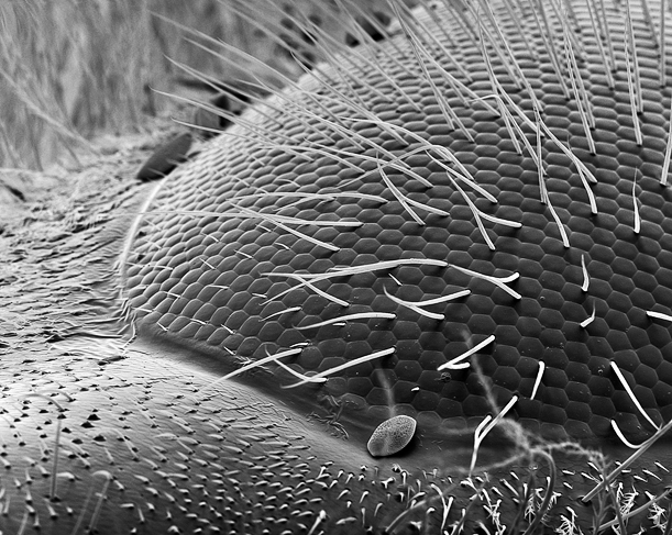
An eye, magnified 190 times. Photo © Rose-Lynn Fisher, Courtesy of th artist and the Craig Krull Gallery, Santa Monica, CA
When Fisher first looked at the creature through the device, she was awestruck by the structures that comprised its body at scales naked to the human eye. One of the first that captured her attention was the bee’s multi-lensed compound eye. “In that first moment, when I saw its eye, I realized that the bees’ eyes are composed of hexagons, which echo the structure of the honeycomb,” she says. “I stood there, just thinking about that, and how there are these geometrical patterns in nature that just keep on repeating themselves.”
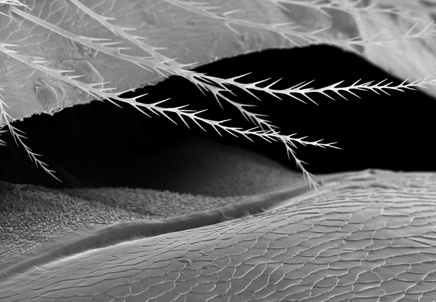
The folded terrain of a bee’s abdomen, magnified 370 times. Photo © Rose-Lynn Fisher, Courtesy of the artist and the Craig Krull Gallery, Santa Monica, CA
Fisher was inspired to continue exploring the body of that bee, and others, continually looking at their microscopic structures and organs in greater and greater detail.
Her creative process started with the obvious: collecting a specimen to examine. “First, I’d find a bee, and look at it through my own regular light microscope to confirm its parts were intact,” she says. “The freshest ones were the best, so sometimes I’d find one walking on the ground that looked like it wouldn’t be around much longer, and I’d bring it home and feed it some honey, to give it something nice for its last meal.” Some of these were rejuvenated by her care, but those that weren’t, and perished, became the subjects of her microscopic exploration.
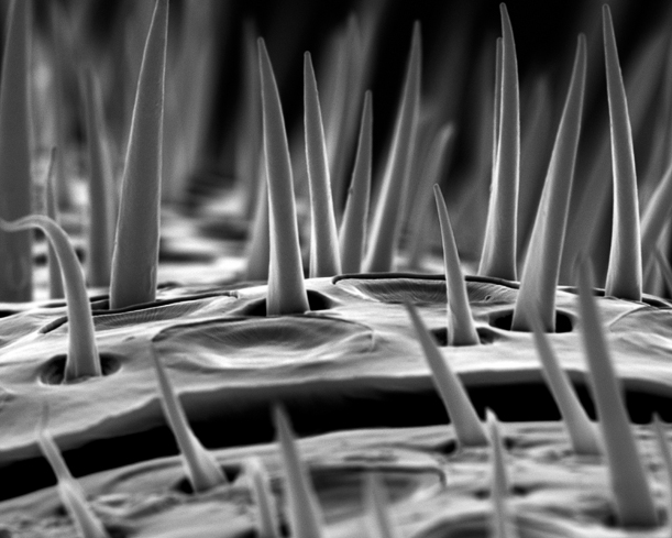
A bee’s microantennae, magnified 3300 times. Photo © Rose-Lynn Fisher, Courtesy of the artist and the Craig Krull Gallery, Santa Monica, CA
At her friend’s lab, in off hours, Fisher used a model of scanning electron microscope called a JEOL 6100, which can detect objects as small as 40 angstroms (for comparison, a thin human hair is roughly 500,000 angstroms in diameter). Before scanning, she’d carefully coat the bee in an ultra-thin layer of gold sputter coating.
This coating, she explains, enhanced the electrical conductivity of the bee’s surfaces, which allow the microscope to detect them in finer resolution. “The SEM uses a very finely focused electron beam that scans across the surface of the prepared sample,” she says. ‘It’s akin to shining a flashlight across the surface of an object in a dark room, which articulates the form with light. With an SEM, it’s electrons, not light—as it moves across the bee’s surface, it’s converting electrical signals into a viewable image.”

The joint between a bee’s wing and body, magnified 550 times. Photo © Rose-Lynn Fisher, Courtesy of the artist and the Craig Krull Gallery, Santa Monica, CA
Once the bee specimen was prepared and mounted inside the SEM’s vacuum chamber, Fisher could use the machine to view the insect at different angles, and manipulated the magnification to look for interesting images. At times, zooming in on the structures abstracted them beyond recognition, or yielded surprising views she’d never thought she’d see looking at a bee.
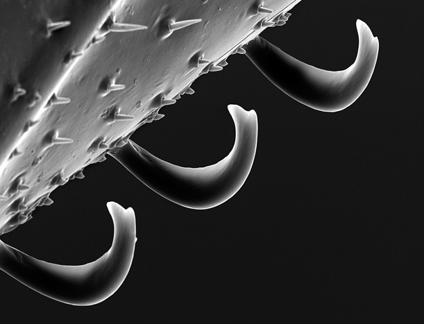
The hooks that attach the forewing and hindwing, magnified 700 times. Photo © Rose-Lynn Fisher, Courtesy of the artist and hte Craig Krull Gallery, Santa Monica, CA
“For instance, when I looked at the attachment between the wing and the forewing, I saw these hooks,” she says. “When I magnified them 700 times, their structure was amazing. They just looked so industrial.”
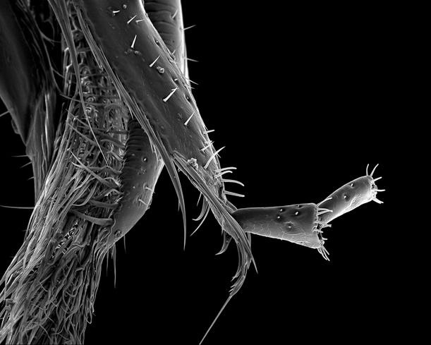
A proboscis, magnified 150 times. Photo © Rose-Lynn Fisher, Courtesy of the artist and the Craig Krull Gallery, Santa Monica, CA
Zoom in close enough, she found, and a bee stops looking anything like a bee—its exoskeleton resembles a desert landscape, and its proboscis looks like some piece of futuristic machinery from a sci-fi movie. At times, Fisher says, “you can go in deeper and deeper, and at at a certain level, your whole sense of scale gets confounded. It becomes hard to tell whether you’re observing something from very close up, or from very far away.”
For more beautiful bee art, see Sam Droege’s bee portraits shot for the U.S. Geological Survey
/https://tf-cmsv2-smithsonianmag-media.s3.amazonaws.com/accounts/headshot/joseph-stromberg-240.jpg)
/https://tf-cmsv2-smithsonianmag-media.s3.amazonaws.com/accounts/headshot/joseph-stromberg-240.jpg)