Revel in the Big Details of Tiny Things With These Prize-Winning Images
Skin cells, tape worms and fuzzy mold are among this years top photos
An entire world exists beyond what the naked eye can see—neural networks snake throughout your body, viruses and bacteria wriggle across tabletops, scales form neat rows atop the wings of butterflies. This fantastical world is brought to light each year in Nikon's Small World Photography Competition. And this year's winners do not disappoint.
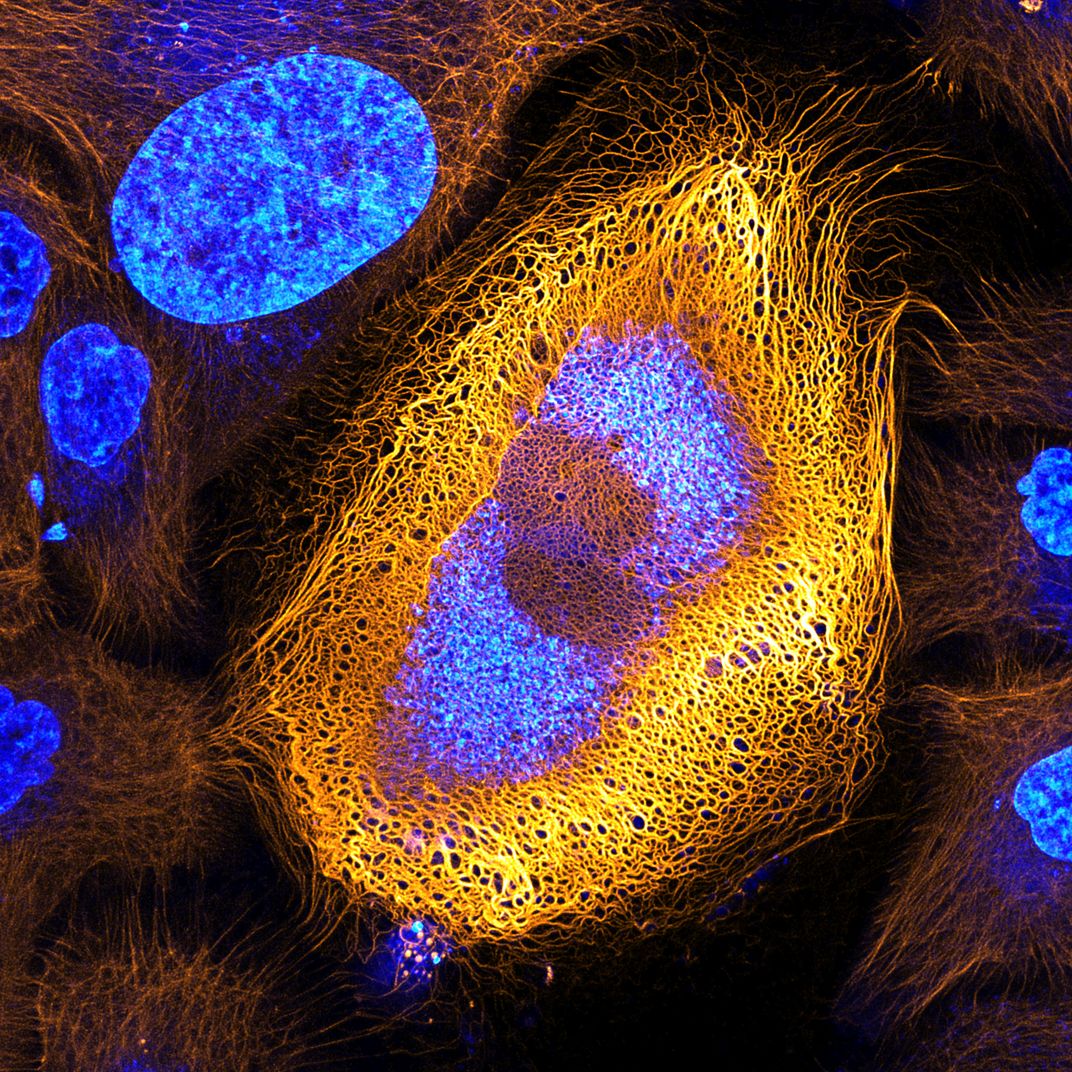
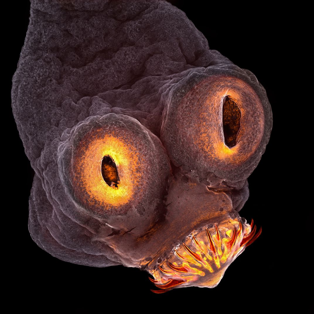
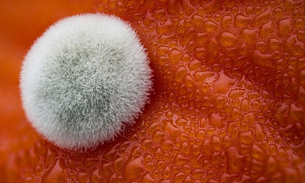
Now in its 43rd year, the contest calls for entries that showcase "the beauty and complexity of life as seen through the light microscope," according to its website. A panel judges, including science researchers and science communicators, selected this year's winning images from a pool of more than 2,000 entries from 88 countries around the world. The ghastly glow of a tapeworm lights up one image, the fuzz of mold emerges from a tomato in another. But the grand prize winner of this year's contest is the wiry network of keratin in a skin cell.
The winning photographer, Bram van den Broek, spends much of his time staring at the world just out of sight as a researcher with the Netherlands Cancer Institute. He captured the prize-winning image while studying how filaments of keratin—a protein found in human skin, hair, nails and more—changes over time in skin cells. To visualize the keratin, he marks it with a fluorescent tag, causing it to glow. The winning image captures one particular cell that caught van den Broek's eye, displaying an excessive amount of the protein, which shows up bold and bright against the darkness of the surrounding cells.
For van den Broek, scrutinizing the complex wiring in skin cells is about more than just capturing compelling images. Rather, it can actually help diagnose and treat skin cancers before they turn deadly. "The expression patterns of keratin are often abnormal in skin tumor cells, and it is thus widely used as tumor marker in cancer diagnostics," van den Broek says in a statement. "By studying the ways different proteins like keratin dynamically change within a cell, we can better understand the progression of cancers and other diseases."
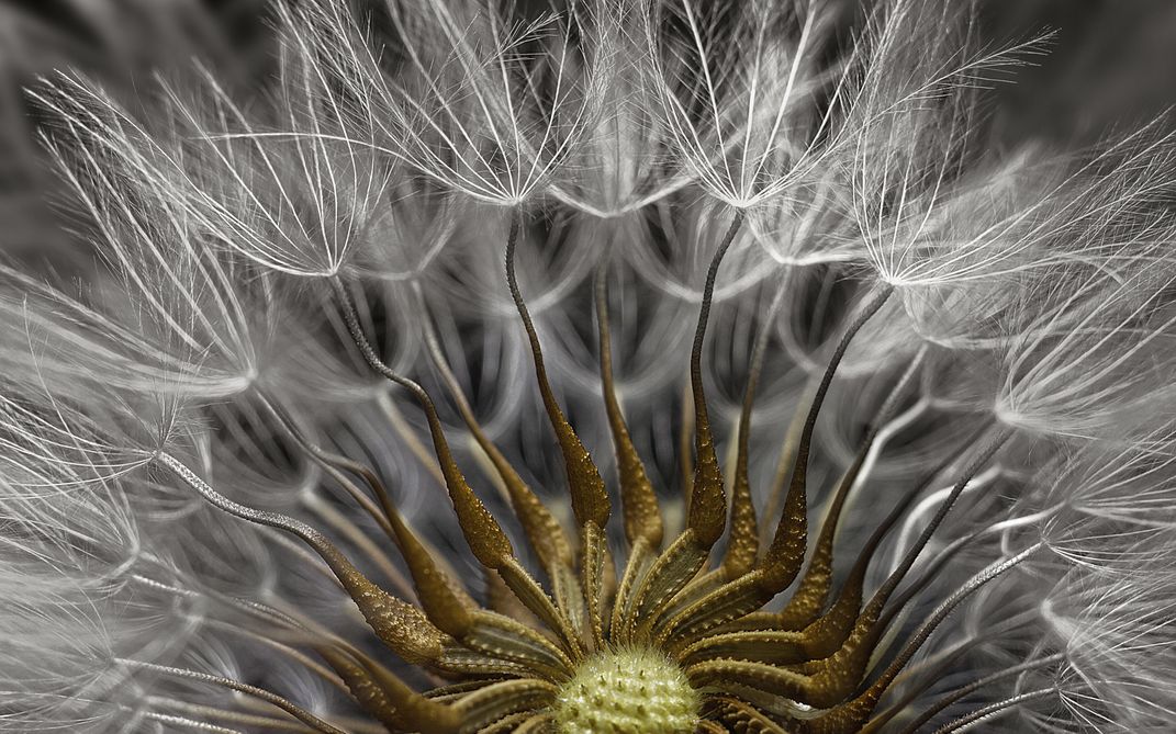
This year's other winning images are equally as captivating. Spikes and fibers protrude from the flowering head of a herb groundsel in Havi Sarfaty's second-place image. A veterinary ophthalmologist, Sarfaty became interested in photomicrography while conducting surgeries under microscopes. The winning image displays something we may see every day in an entirely new light.
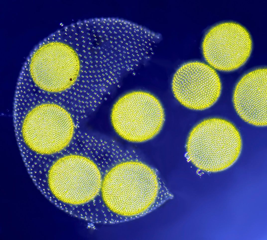
The third-place image highlights a mature colony of volvox algae, a type of tiny greenery that commonly grows in fresh water. The globular colony is frozen in mid-rupture, releasing its brightly colored daughter colonies into the world to reproduce. The photographer that captured this image, Jean-Marc Babalian, has been taking photographs of the microscopic world for three decades. He's a production manager for a French construction materials company.
Take a spin through more of the microscopic world by viewing the rest of the images on the competition's website. And perhaps next year you too can join in the fun of seeking what lies just beyond what your eyes can see.
