See Microscopic Butterfly Wing Scales Materialize Inside of a Chrysalis
The study is the most detailed look at the structures to date and could be used to design new materials
:focal(450x300:451x301)/https://tf-cmsv2-smithsonianmag-media.s3.amazonaws.com/filer_public/bd/dc/bddc588e-7746-43e1-bbdc-5d6f6ce387dc/mit-butterfly-growth-01-press_0.jpg)
Butterflies are adored for their diverse wing patterning and metallic colors. The shimmering hues come from a meticulous arrangement of thousands of microscopic scales on their wings. These tiny structures give the insects protection against the elements and stabilize their body temperatures.
Now, scientists at Massachusetts Institute of Technology (MIT) have developed a way to peek inside of a butterfly's chrysalis and record in real-time how these scales develop from start to finish, reports Hannah Seo for Popular Science. The study was published this month in the Proceedings of the National Academy of Sciences.
The iridescence on butterfly wings does not occur from pigment molecules but by how the butterfly wing is structured. Physicists call it photonic crystals, a term that can be used to describe the common iridescent effect seen on many other insect wings and even opals. A butterfly wing's shimmering qualities materialize when a versatile molecule called chitin forms scales arranged like roof tiles, reports Jennifer Ouellette for Ars Technica. The arrangement splits and diffracts light into several beams in different directions in an optical concept known as diffraction grating. Another example of this phenomenon is seen in the dancing waves of light seen on the reflective side of a CD. However, photonic crystals only reflect specific colors or certain wavelengths of light, which give butterflies their unique coloration. Diffraction grating alone will reflect the entire spectrum of color, but adds iridescence when accompanied by photonic crystals, Ars Technica reports.
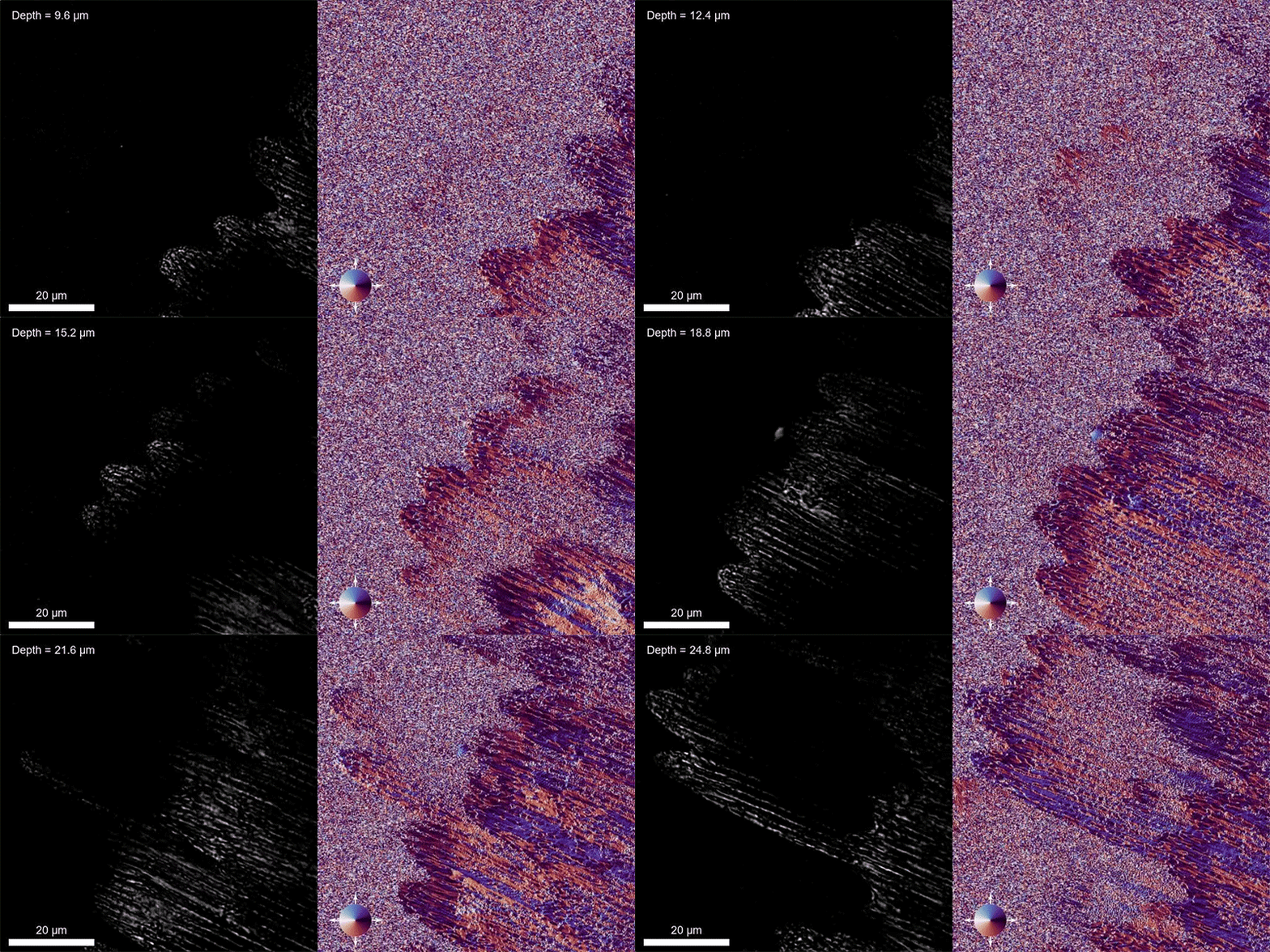
To image wing formation inside the chrysalis, researchers raised groups of painted lady butterflies (Vanessa carduli). They waited until the caterpillars began their transformation inside the chrysalis and then sliced the cuticle open to create a viewing window. Per Popular Science, the team then covered the opening with a small piece of glass called a coverslip. Researchers imaged and recorded the development of each insect's hindwing and forewing using this process.

Viewing the wings using a standard beam of light would have damaged the cells. To record the wing's formation process without damaging the delicate cells, the research team used speckle-correlation reflection phase microscopy. This type of microscopy works by shining tiny points of light onto a specific area on the wing, Ars Technica reports.
"A speckled field is like thousands of fireflies that generate a field of illumination points," Peter So, an imaging expert at MIT and one of the study's collaborators, said in a statement. "Using this method, we can isolate the light coming from different layers, and can reconstruct the information to map efficiently a structure in 3-D."
In the team's video footage, they found that cells started lining up in rows along the wings' structure within days that metamorphosis began. After initially lining up, the cells began to differentiate themselves in an alternating pattern of cover scales overlaying the wing and ground scales that grew underneath the wing, per Popular Science. Researchers expected to see the cells wrinkle and compress in the final growth step. Instead, they developed a wavy, ridged structure.
The team plans on further exploring the structure of butterfly wings and the reasoning behind the ridged design. Unlocking the methods behind butterfly scale formation could lead to bioinspired technology like new solar cells, optical sensors or rain- and heat-resistant surfaces. Another application could be iridescent encrypted currency to discourage counterfeiting, per a statement.
/https://tf-cmsv2-smithsonianmag-media.s3.amazonaws.com/accounts/headshot/gamillo007710829-005_0.png)
/https://tf-cmsv2-smithsonianmag-media.s3.amazonaws.com/accounts/headshot/gamillo007710829-005_0.png)